Research Review Corner
ARPA-H, which stands for Advanced Research Projects Agency for Health, is an agency within the U.S. Department of Health and Human Services. One of its major projects is called the FRONT program which stands for Functional Repair of Neocortical Tissue . The FRONT program focuses on repair and regeneration of the neocortex, the largest part of the brain. The neocortex is critical for many aspects of cognition and everyday functioning. The neocortex can be damaged in many ways including stroke, traumatic injury, and neurodegeneration, such as in Alzheimer's disease. Serious damage to the neocortex often causes individuals to be dependent on costly therapies or caregivers and can also lead to related health and medical problems. The FRONT program hopes to alter this pattern by using cutting-edge neurodevelopmental principles and stem cell technology to regenerate brain tissue and restore lost functions.
For decades, families facing severe traumatic brain injury have lived with a painful assumption: once brain tissue is lost, recovery is limited to what the surviving brain can re-learn. Rehabilitation can help. Technology can assist. Time can produce surprises. But the central wound—the loss of brain cells, circuits, and connections—has often been treated as permanent. That assumption is beginning to change.
A Neuroengineer’s Approach to Rebuilding Neural Circuits Note: Justin Burrell is the 2025 winner of the DLF Prize for Post-Doctoral Research. This article emphasizes the regenerative potential of improving the connectivity of replenished neural material.
For most of modern medical history, the brain has been viewed as incapable of regeneration. While skin, bone, and even parts of the liver can regenerate after injury, damage to the brain—whether due to stroke, traumatic brain injury (TBI), or neurodegenerative disease—has long been considered largely irreversible. Over the past decade, however, advances across stem-cell biology, neuroengineering, and computational neuroscience are challenging this dogma. Today, a broad set of scientific strategies is aimed at enabling true repair of damaged neural circuits . Many of these scientific strategies have been highlighted in previous editions of the DLF newsletter Neural Connections ( archived at the DLF website ). Although each approach faces obstacles, the collective progress is significant enough to shift expectations about what may one day be possible. This article focuses on one strategy that has produced some of the most dramatic and tangible preclinical results: transplantation of human stem-cell–derived neural tissue.

On July 10, 2025, the Advanced Research Projects Agency for Health (ARPA-H) announced a major initiative titled Functional Repair of Neocortical Tissue or FRONT. The announcement states “FRONT will pioneer a curative therapy for the more than 20 million adults in the US living with chronic neocortical brain damage from neurodegeneration, stroke, trauma, and other causes, which costs the country an estimated $800 billion per year. Worldwide, more than 200 million people live with debilitating after-effects of brain damage.” A set of informational meetings about this program and a due date for outlines of potential proposals have been set for August. Full proposals are due by September 25, 2025. Complete instructions, specifications, and expectations are delineated in the ARPA-H FRONT announcement. The FRONT announcement includes a clear expectation that the successful brain regeneration methods that are discovered will be used in clinical trials with persons with brain injury by the fifth year of the program. The DLF lauds ARPA-H for initiating this program. We are discussing possibilities for playing a supportive role as proposals develop. This exciting program is congruent with the original overarching goals of the DLF and confirms the validity of its mission.
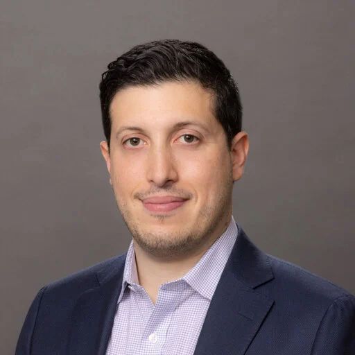
Dr. Burrell is a translational neuroengineer in the Departments of Neurosurgery and Oral & Maxillofacial Surgery at the University of Pennsylvania. His research integrates advanced neural repair strategies with clinical translation, focusing on axon protection, nerve fusion, and engineered neural tissue for neurotrauma recovery. Dr. Burrell has led the development of multiple first-in-field innovations—including the first large-animal model of nerve fusion, delayed axonal fusion protocols, and the first orally active axonal protectants—positioning him as a recognized leader in regenerative neurotechnologies. He is co-founder of Neurostorative LLC and plays a central role in several other platforms aimed at neural reconnection, long-term preservation, and bio-integrated prosthetic systems.
The human brain is remarkable in its complexity, adaptability, and resilience. Yet, millions worldwide face dramatically impaired quality of life due to traumatic brain injuries, strokes, and degenerative neurological diseases. Today, there are no medicines that stimulate the restoration of lost brain functions. Recent research is showing us a path to new medicines that may activate the brain’s inherent capacity for regeneration following severe injury. One notable researcher in this pioneering field is Jared Tangeman at Johns Hopkins University. Tangeman investigates the extraordinary regenerative capabilities of the axolotl, a salamander species uniquely capable of completely regenerating its central nervous system (CNS), including brain and spinal cord tissues. Through sophisticated genetic and cellular analyses, his lab has uncovered essential genetic programs activated during neural regeneration. Tangeman’s laboratory employs innovative models of optic nerve injuries, such as whole-eye enucleation (temporary removal and reimplantation of the entire eye in the salamander) to explore how retinal cells survive extreme conditions and regenerate nerve connections. Utilizing advanced genomic techniques, including single-cell RNA sequencing and chromatin profiling, paired with AI-driven structural modeling, Tangeman identifies critical genes such as ATF3 and RUNX1, which regulate neuronal survival, axon regeneration, and the activation of progenitor cells into new neurons¹. Identifying specific genes involved in regeneration provides precise molecular targets for developing new therapeutic approaches. Once these targets are validated in human cell-based models, they can guide the development of novel medicines, particularly genomically targeted drugs, to enable human brain regeneration after severe injury. Complementing Tangeman’s fundamental research is work led by Justin Burrell at the University of Pennsylvania. Burrell is focused on rapidly translating laboratory findings into clinically viable therapies. His team discovered boldine, a compound with remarkable neuroprotective effects. This compound has been shown to reduce nerve damage and promote nerve regeneration in animal studies². These findings have potential implications for treating nerve injuries and TBI. Additionally, Burrell’s team has developed engineered neural scaffolds, biomaterials designed to guide nerve fibers across injury sites, which have successfully reconnected severed nerves in preclinical studies³. These tissue-engineered constructs, when combined with pharmacological interventions such as boldine, offer promising dual-action treatments capable of dramatically improving recovery outcomes for severe brain injuries. The innovative research by Tangeman and Burrell showcases a powerful synergy—Tangeman’s foundational insights into molecular regeneration mechanisms provide precise targets for novel therapies, while Burrell’s work demonstrates practical strategies immediately applicable in clinical settings. A promising therapeutic frontier inspired by these fundamental discoveries involves genomically targeted medicines such as antisense oligonucleotides (ASOs) and RNA interference (RNAi) molecules. ASOs are short, synthetic molecules designed to precisely modulate gene expression by targeting specific RNA sequences, thus influencing cellular behaviors and disease processes⁴. ASOs have demonstrated clinical efficacy in treating genetic diseases, including spinal muscular atrophy, underscoring their therapeutic potential for neurological conditions⁵. Similarly, RNAi-based therapies, designed to silence specific genes involved in disease progression, have recently entered clinical practice for conditions such as hereditary amyloidosis, further underscoring their potential⁶. Notably, ASO-based therapies are currently being tested in clinical trials for spinal cord injuries, aiming to harness the CNS’s innate ability to restructure itself. These pioneering trials, led by neuroscientists such as Stephen Strittmatter at Yale University, indicate a viable pathway toward applying similar strategies to brain injuries⁷. The convergence of Tangeman’s foundational genetic discoveries and Burrell’s translational approaches positions genomically targeted therapies, including ASOs and RNAi, as highly promising tools for stimulating regeneration and recovery. Once validated in human cell-based models, these targeted genomic strategies can significantly enhance regenerative capacities in human brains after severe injury. Concurrent advances in biomaterials, nanomedicine, and artificial intelligence (AI) further bolster regenerative research. For example, nanotechnologies enable the targeted delivery of therapeutics directly to injured brain regions, increasing efficacy and precision⁸. AI-driven tools rapidly analyze complex genetic and molecular data, swiftly identifying promising targets and therapeutic strategies. Collectively, the groundbreaking efforts of researchers like Jared Tangeman and Justin Burrell, alongside advances in genomically targeted therapies, nanomedicine, and AI, hold transformative potential. These innovations significantly advance our understanding and ability to promote neural regeneration, offering renewed hope and improved quality of life for millions affected by severe neurological injuries.
Introduction: The Paradigm Shift in Brain Regeneration For decades, scientists believed that the adult brain was incapable of meaningful regeneration. Unlike skin or muscle, the central nervous system (CNS) appeared to lack the ability to repair itself after injury, and lost neurons were considered irreplaceable. However, cutting-edge research now demonstrates that this is no longer the case. The brain possesses dormant repair mechanisms—pathways that were active during development but have been shut down in adulthood. By reactivating these pathways, it may be possible to induce neurogenesis (the birth of new neurons), axonal repair, and synaptogenesis (the formation of new neuronal connections) after devastating injuries like stroke, traumatic brain injury, and spinal cord damage. A growing body of evidence from both the spinal cord and the central nervous system (the CNS) shows that regeneration can be stimulated by downregulating the repressing factors that prevent neuron growth and repair. For clarity, “downregulation” means reducing the activity or number of something. In the case of a repressing factor, downregulation means lowering its levels or making it less active. A repressor is a protein that blocks a process—often by preventing a gene from being turned on. When the repressor is downregulated, it no longer effectively blocks that process, allowing the underlying function to be released or activated. These advances open the door to new treatments that could restore function to patients with neurological injuries, potentially reversing what were once thought to be permanent disabilities. The Science Behind Reactivating Brain Repair Neurodevelopmental genes and pathways that promote neuronal growth and plasticity are switched off after early development. However, researchers have identified key molecular regulators that act as “brakes” on brain regeneration. The “brakes” act as repressors of brain regeneration. By inhibiting these repressors, the brain’s intrinsic ability to regrow neurons and axons can be reactivated. Recent breakthroughs include: Neurogenesis in the Adult Brain : Research has shown that downregulation of PTBP1, an RNA-binding protein, can convert glial cells into neurons in the adult brain. Studies using antisense oligonucleotide (ASO) therapy to transiently suppress PTBP1 have successfully induced the formation of functional neurons in mouse models of neurodegeneration. For clarity, an antisense oligonucleotide (ASO) is a short strand of DNA or RNA designed to bind to a specific mRNA and regulate gene expression. By binding to its target, an ASO can block protein production, modify splicing, or mark the mRNA for destruction, effectively acting as a precise “off switch” for genes. In this context ASOs can be used to release glial cells to develop into functional neurons. Work is now underway to create ASOs that can be safely administered to humans, thereby stimulating the creation of new neurons. Axonal Regeneration in the CNS: Another major discovery involves unlocking nerve fiber regrowth by downregulating specific nerve growth inhibition, which prevents nerve fiber regrowth. Studies have demonstrated that suppressing these inhibitors leads to long-distance axonal regeneration, restoring function in spinal cord injury models. Synaptic Repair in Neurodegenerative Disease: Scientists have shown that modulating key synaptic receptors—such as mGluR5—can restore lost synaptic connectivity, improving brain network function in neurodegenerative diseases like Alzheimer’s. The neuronal connections and circuits in the brain are maintained in a stable state by the balanced action of different synaptic receptors. By modifying this balance, the rate of brain synapse formation can be increased or decreased. Together, these findings confirm that neural repair is possible when the appropriate repressive factors are removed, unlocking the brain’s natural regenerative capacity. Quiver’s Scalable Human Neuronal Platform: Accelerating Discovery While these discoveries are promising, translating them into effective therapies requires precise, scalable, and high-throughput screening technologies that can screen out or discover biomolecules that address a specific biomedical target by the thousands with great rapidity. One such technology has been developed by Quiver Biosciences using an all-optical electrophysiology platform. (Note: the author of this article is a co-founder of Quiver Biosciences) Quiver has developed a human induced pluripotent stem cell (hIPSC)-derived neuronal platform that allows researchers to measure functional activity in human neurons at an unprecedented scale. This platform is uniquely capable of: Directly measuring neuronal excitability, synaptic transmission, and network connectivity using optogenetics and advanced imaging techniques. Screening for new factors that inhibit brain repair, helping scientists identify additional repressors that need to be targeted. Evaluating small molecules and ASO-based therapies that may upregulate brain regeneration, assessing their efficacy and toxicity before advancing to clinical trials. The potential of this platform is vast. Scientists can now systematically search for drugs that mimic the effects of PTBP1 suppression, Nogo-A blockade, or mGluR5modulation—treatments that could one day be used to regrow neurons, reconnect severed axons, and restore lost synapses in patients with brain injuries. Key advantages of Quiver’s platform include: Scalability: Unlike traditional animal models, this system enables high- throughput drug screening on human neurons, increasing the efficiency of therapeutic discovery. Relevance: Because the neurons are derived from human stem cells, they offer a more accurate model of human brain function and disease than animal-based approaches. Safety Screening: The platform allows for early identification of toxic effects, ensuring that only the safest and most effective compounds move forward in development. By integrating AI and machine learning, Quiver’s platform also enables pattern recognition of successful drug candidates, identifying compounds with optimal efficacy and minimal side effects. The Role of Philanthropy: Funding the Future of Brain Repair Scientific breakthroughs alone are not enough to bring regenerative therapies to patients. Translational research—the process of developing basic discoveries into real-world treatments—is slow, expensive, and underfunded. This is where philanthropic support can make an immediate impact. Many of the pioneering studies on brain regeneration were initially considered too risky or unconventional for traditional funding sources. Yet, thanks to early philanthropic investments, these ideas have now been validated and are shaping the next generation of treatments. Today, funding is urgently needed to: Expand screening for regeneration-promoting drugs Optimize ASO-based approaches for inducing neurogenesis and axonal repair. Accelerate clinical trials for promising regenerative therapies. A New Era for Brain Regeneration For the first time, we have the tools to reactivate the brain’s own repair mechanisms, offering hope for individuals with severe neurological injuries. The path forward is clear: by investing in innovative research, scaling up discovery efforts, and supporting translational studies, we can bring brain-regenerating therapies to patients faster. The DLF raises funds and uses them to inspire, catalyze, and accelerate work towards brain regeneration. We increase public awareness of possibilities in brain regeneration through our social media, news “blasts”, and quarterly newsletter. And, we promote neuroscientific advances through consultation, networking, conferences, and seed grants. This is especially important when federal funding is limited. By supporting this work, we can help transform groundbreaking scientific insights into real treatments that restore lost function and improve lives.
Stroke is a common neurological condition that damages brain cells (neurons) in the affected area, leading to a loss of the functions controlled by that region. A hopeful aspect of stroke recovery is that, over time and with rehabilitation, many individuals regain some abilities. This recovery has been linked to a process called “remapping,” where neurons in unaffected areas of the brain adapt to take over the functions of the damaged areas. Although many studies have explored this remapping phenomenon, most evidence has been indirect, based on changes in brain activation patterns or neuron connections after stroke in animal models. Direct proof that neurons change functionality after stroke has been lacking, partly because measuring neuron activity in the brain over time, especially at the necessary scale and duration, is challenging.
Scientists worldwide are working to find ways to stimulate healing and functional recovery after severe brain injuries. This work is driven by the desperate needs of persons who have suffered brain damage. It is inspired by the knowledge that the information required to create new brain cells, cause these cells to interconnect, and stimulate new learning is contained in our genome. Now that we can readily generate stem cells from adult tissue, we have access to the genomic program that can control all of the intricate details of brain tissue formation. A number of different research themes are being pursued productively. These include: (1) enabling injured neurons to self-repair (“axonal repair”) 1,2 ; (2) replacing damaged tissue by increasing the growth of new neurons (“neurogenesis”) 3-5 ; (3) transplanting new brain cells that are derived from a person’s own stem cells (“autologous cellular repletion”) 6-8 ; (4) stimulating the re-wiring of new or surviving tissue by encouraging the formation of new connections (“synaptogenesis”) 9,10 ; and (5) augmenting the function of a damaged brain by the use of bio-computational prostheses (“brain-computer interfaces”) 11,12 ; We’ve explored these themes in previous newsletters. The goal of stimulating meaningful brain regeneration is now sufficiently plausible that a large-scale, well-funded campaign needs to be funded to bring meaningful new therapies to patients within the foreseeable future. Here, we suggest a high-level outline of the research themes for such a campaign. A ‘moon shot’ program towards brain regeneration would leverage cutting-edge technologies in stem cell research, gene therapy, synaptic plasticity, neuronal repair, and brain-computer interfaces (BCIs) to develop innovative treatments for brain injuries and neurodegenerative diseases. These treatments would target the restoration of lost brain functions and improvement in the quality of life for individuals affected by severe brain injuries. This research agenda aims to catalyze serious discussion about creating a federal program with funding, organizational resources, and expert governance to enable brain regeneration in our lifetimes. Major Themes For a Brain Regeneration “Moon Shot” Program 1: Promote the formation of new neurons 1.1 Stimulate the brain to create new neurons 1.2 Create new neurons from patient-derived induced pluripotent stem cells to be transplanted back into the patient. Create new glial cells to support neurogenesis. 2: Stimulate new synaptic formation 2.1 Develop drugs that enhance synaptic plasticity and promote the formation of new synaptic connections 3: Stimulate self-repair of damaged neurons 3.1 Develop drugs that de-repress neurons and, thereby, enable axonal regrowth 4: Develop brain-computer interfaces (BCIs) for brain-injured patients 4.1: Develop and test BCIs that enable the brain to control behaviors or external devices and, thereby, augment or replace impaired functions. 4.2: Develop and test BCIs that can accelerate the training of remapped brain tissue in persons with brain injuries to optimize functional recovery. 4.3: Combine BCIs with other strategies (e.g., cell repletion, synaptogenesis, and enhanced plasticity) to accelerate adaptation and functional improvement. The proposed research themes can underpin targeted research to stimulate meaningful brain regeneration, offering new hope for patients with brain injuries and neurodegenerative diseases. While the scientific challenges are profound, there has been sufficient progress to justify substantial investment in brain regeneration research. Any such large-scale program will require coordinated collaborations among academic and commercial partners, skillful governance and management, and a shared sense of profound commitment to the goal. The recent pace of advances in cell biology, stem cell technology, bio-computational interfaces, and genomically targeting medicines suggests that large-scale investment will yield meaningful clinical advances toward brain regeneration after injury. With robust funding and skilled leadership, this comprehensive research agenda has a realistic potential to transform scientific breakthroughs into tangible medical therapies, offering hope to millions affected by brain damage.